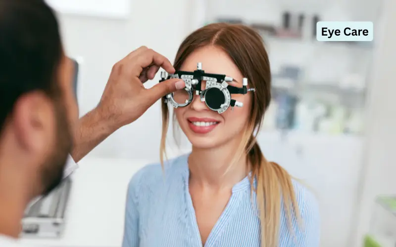
Noticing subtle visual fatigue after long screen sessions feels a lot like ignoring minor performance degradation in a core production service. When your focus flickers or contrast softens by late afternoon, booking a quick glasses update feels like the easiest fix, but optical lenses only adjust incoming light without inspecting the neural sensors underneath.
Protecting your long-term sight requires examining deep retinal micro-structures and macular thickness rather than simply swapping lens prescriptions. If you are scheduling a comprehensive eye health audit with an eye specialist in Singapore, obtaining high-definition optical coherence tomography scans gives you an objective baseline for tracking your ocular foundation over time. Establishing a proactive diagnostic roadmap early protects your central vision before irreversible nerve cell loss takes place.
The Hidden Strain of High Myopia
High nearsightedness is far more than an inconvenience that requires thick spectacle lenses; it fundamentally alters the mechanical shape of your eyeball. When an eye grows longer from front to back, the delicate retinal tissue lining the posterior wall stretches and thins across the ocular curve.
Because the retina lacks sensory pain fibres, progressive thinning, lattice degeneration, and microscopic breaks occur without any physical discomfort. Over several years, mechanical traction from the shrinking vitreous gel can pull on these stretched zones, creating tiny tears that allow intraocular fluid to seep behind the retina, silently threatening central vision.
Surface Fundus Photos versus Deep Scans
Standard high-street optometry chains frequently offer basic digital fundus photography as a complete retinal screening. The non-obvious trade-off is that two-dimensional fundus photos capture only the surface colour of the retina, leaving the ten microscopic cellular layers underneath completely invisible.
Choosing optical coherence tomography trades a few extra minutes in the clinic chair for cross-sectional cellular imaging. Much like an ultrasound that uses light waves, this advanced scan slices through tissue layers down to the micron level. It detects early sub-macular fluid accumulation, occult choroidal neovascularisation, and microscopic vitreoretinal traction years before standard fundus photography shows any visible abnormality.

The Prescription Update Trap
The most common and expensive mistake working professionals make is treating progressive reading difficulty solely as an optical refraction issue. People fall into this trap because buying a stronger pair of reading spectacles temporarily sharpens text, creating the false impression that their eye health is perfectly sound.
In reality, underlying macular degeneration and diabetic micro-aneurysms can develop in parallel with routine presbyopia. Relying on successive spectacle updates without dilating the pupil masks silent vascular leaks and geographic atrophy. By the time central vision becomes permanently distorted, the opportunity for early, non-invasive laser seals or anti-VEGF injections has already passed, leaving patients with irreversible blind spots.
Calculating Five Year Preventative Care Economics
Let us examine a concrete worked example comparing reactive emergency vitreoretinal intervention against proactive diagnostic monitoring over five years. Consider an individual with moderate myopia who skips detailed clinical retinal screenings to save money.
When an unmonitored peripheral tear progresses into a subtotal retinal detachment, emergency surgical vitrectomy, gas tamponade, and post-operative monitoring accumulate 14,800 dollars, alongside four weeks of lost professional billing worth 12,000 dollars, totalling 26,800 dollars. In contrast, an initial specialist clinical screening costing 380 dollars identifies the peripheral tear early, enabling a ten-minute laser retinopexy costing 1,400 dollars that requires zero time away from work. Sourcing a proactive diagnostic pathway saves 25,020 dollars while preserving pristine visual clarity.
Practical Daily Habits for Retina Protection
Maintaining optimal retinal circulation throughout decades of intense digital work requires establishing simple, repeatable operational habits. Treating your eyes like high-precision optical hardware keeps metabolic tissue stress low.
Incorporate these practical operational habits into your daily routine:
- Follow the twenty-twenty distance break: Look away from digital monitors at an object twenty feet away for twenty seconds every twenty minutes to ease ciliary muscle spasm.
- Wear polarised ultraviolet sunglasses outdoors: Protect delicate macular pigments from high-energy blue and ultraviolet radiation that accelerates retinal photo-oxidation.
- Consume dietary carotenoid antioxidants daily: Include lutein and zeaxanthin from leafy greens, eggs, and bright vegetables to reinforce protective macular pigment density.
A Simple Grid Rule for Distortions
You do not need specialised ophthalmic equipment to check your macula between regular clinical appointments. You can apply a straightforward decision rule called the daily straight-edge check using a bathroom tile or window frame.
Cover your left eye with your palm, look directly at a straight horizontal line or an Amsler grid with your right eye, and check for irregularities. If the line appears perfectly straight, crisp, and uniform, your central macular architecture is stable. If any section of that straight line appears bent, wavy, washed out, or missing, do not wait for your annual checkup; contact a specialist clinic within twenty-four hours to inspect for macular fluid accumulation. Repeat the test with your right eye covered to ensure both eyes remain equally sharp.
Preserving your visual acuity for the decades ahead is ultimately about replacing reactive glasses adjustments with structured, proactive ocular monitoring. By looking past surface-level photography, understanding high-myopia tissue physics, and applying clear self-checks, you protect both your career productivity and your long-term independence. Approach your ocular health with thoughtful strategy, rely on solid medical evidence, and move forward with quiet confidence in your personal care decisions.